DDr. D. Zitny-Haberl MDSc | Dr. M. Müller
Pilgramgasse 1/2/8, 1050 Wien, +43 1 5447199
Mon, Tue, Wed, Thu 8am–6pm, Fri 8am–2pm

DDr. D. Zitny-Haberl MDSc
Dr. M. Müller
Pilgramgasse 1/2/8, 1050 Wien
+43 1 5447199
Mon – Thu 8am – 6pm, Fri 8am–2pm

Digital Volume Tomography

»We are now pleased to be able to offer you 3-D, X-ray images! With our Morita, (Veraview X800) X-ray machine we can take outstanding quality images in extremely high resolution.«

Today, pictures of such a high quality are an indispensable tool in modern dentistry, not only diagnostically, but also in root canal and implant surgery. Previously, with 2 dimensional X-ray images, it was often not possible to see enough detail to make an accurate diagnosis.

A digital volume tomography (DVT) makes it possible to display the respective jaw areas in three dimensions. These images display areas on all three levels.

In addition to the many diagnostic advantages of DVT imagery, these digital recordings also enables an accurate measurement of bone density, not only necessary for planning implants, but also to prevent injuries to the lower jaw nerves or other important structures and enables us to make optimal use of the available bone. With this technology, it is possible to plan a computer-controlled implant.

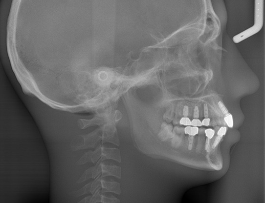
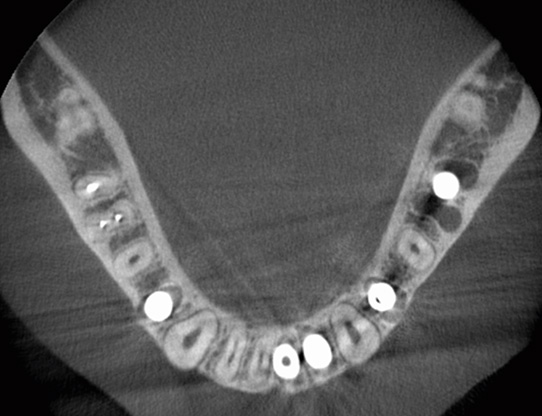
Another important application area of the DVT is also in the optimisation of root canal treatments. Inflammatory changes show up in their early stages, which previously (with conventional X-ray machines) were not yet recognisable. These pictures depict the root canal in three dimensions and clearly shows their positions, which facilitates a perfect root canal filling procedure.

In contrast to conventional CAT scan, the radiation dose is significantly less with a DVT scan.